
Figure 2.
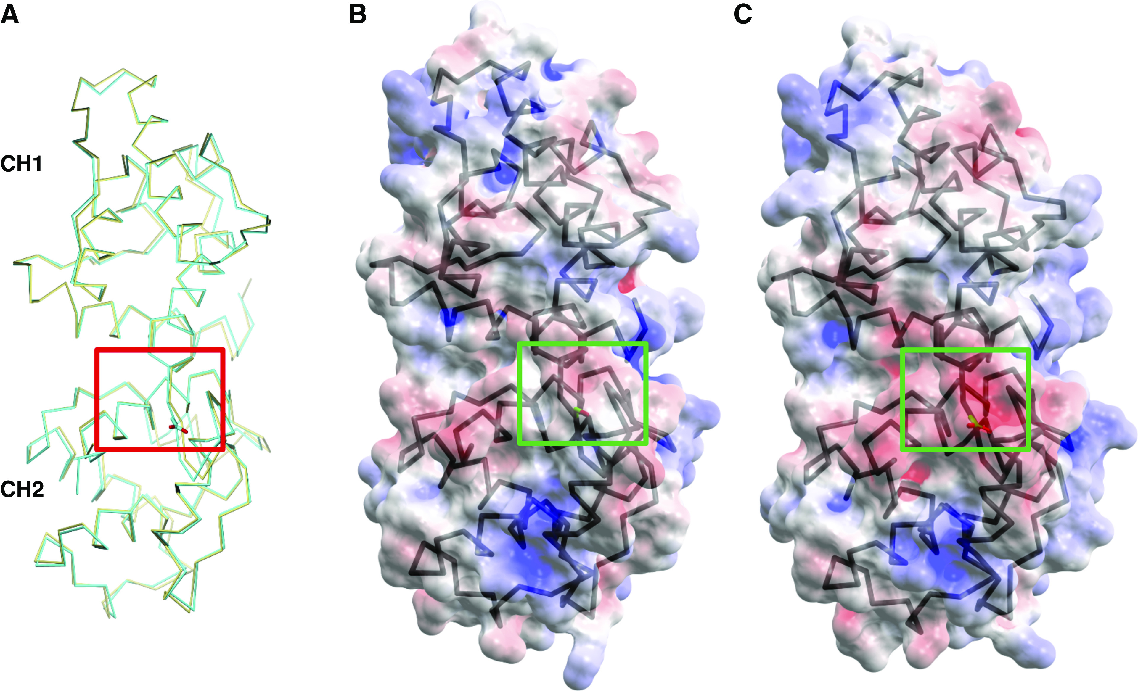
Phosphomimetic S159D ACTN4 does not change the conformation of its ABD. (A) Superposition of the Cα positions of the ABD of WT (khaki) and phosphomimetic S159D (cyan) ACTN4. The red box highlights the side chain of aspartate (D) at position 159. The CH1 domain is shown at the bottom, and the CH2 domain is shown at the top. Semitransparent electrostatic surface representation of (B) WT ACTN4 ABD and (C) phosphomimetic S159D ACTN4 ABD; both are colored according to electrostatic potential (red [acidic, −5 kBT], white [neutral, 0 kBT], and blue [basic, 5 kBT]). Cα positions of the ABD of (B) WT and (C) phosphomimetic S159D ACTN4 are indicated by the black lines. The red box highlights negative charge (represented by red shading) conferred by D159 substitution in (C), which is different from more neutral charge conferred by S159 in (B). Different side chain orientations can be attributed to distinct crystal packing effects.