Table 2.
Suggested abbreviated protocol for hand-held cardiopulmonary ultrasound study in suspected or confirmed COVID patients.
| HH-POCUS Protocol | 2D | Colour flow | Key information |
|---|---|---|---|
 Parasternal Long Axis |
LV and RV Aortic root Pericardium Ascending aorta LA size |
MV AV |
Size (LV, RV) and function (LV, RV, AV, MV) Pericardial and pleural effusion |
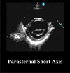 Parasternal Short Axis |
Basal, mid and apical LV AV level and RVOT |
– | Size (LV, RV) and function (LV, RV and AV) |
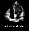 Apical four chamber |
LV and RV LA and RA |
MV TV |
Size (LV and RV, RA, LA) and function (LV and RV, RA, LA, MV and TV). |
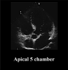 Apical 5 chamber |
AV | AV | Function (AV) |
 Apical two chamber |
LV | MV | Size and function (LV) |
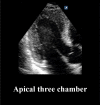 Apical three chamber |
LV AV and MV |
– | Size (LV) and function (LV, RV, AV and MV) |
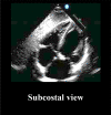 Subcostal view |
Pericardium IVC size and respiratory dynamics |
– | Pericardial effusion RA pressure estimation (IVC size and dynamics) |
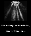 Midaxillary, midclavicular, paravertebral lines |
A or B-lines Pleural line (10 quadrants) |
– | Interstitial syndromes (pulmonary odema, ALI/ARDS) |
Bold text – recommended abbreviated dataset. Normal font – optional images depending on abnormal preceding images of those structures.
