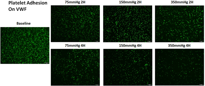
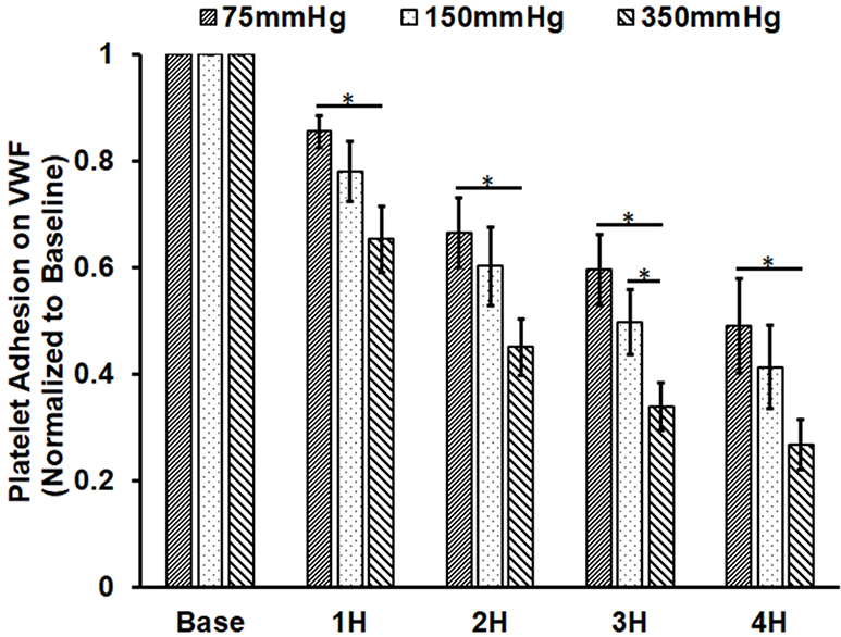
Figure 5.
Altered platelet adhesion on VWF of the blood from the three loops (ΔP=75 mmHg, 150 mmHg and 350 mmHg). A) Representative images of platelet adhesion on VWF from the baseline and two hourly blood samples (the 2nd and 4th hours) (magnification, X400); B) The quantitative comparison of the area coverage of adherent platelets on VWF from the baseline and four hourly samples (*P<0.05).


