Figure 1.

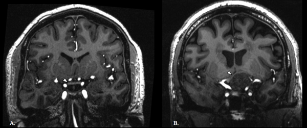
Optic chiasm in control and adenoma patient. Coronal T1-weighted image of a normal optic chiasm (white arrowhead) in a control (A). Coronal T1-weighted image of an optic chiasm (white arrowhead) being compressed by a pituitary adenoma (B).

Optic chiasm in control and adenoma patient. Coronal T1-weighted image of a normal optic chiasm (white arrowhead) in a control (A). Coronal T1-weighted image of an optic chiasm (white arrowhead) being compressed by a pituitary adenoma (B).