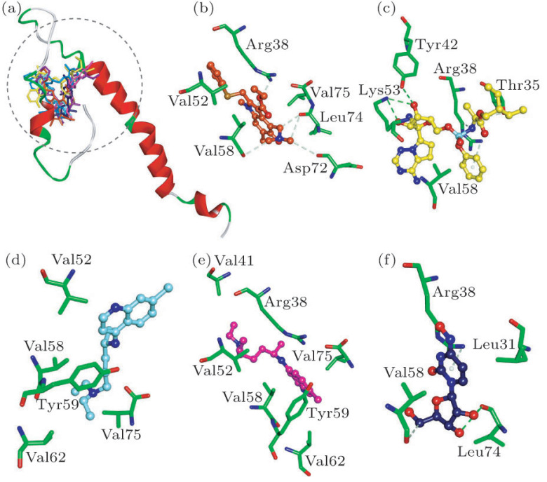
Fig. 1.
(a) Compounds superposed in envelope structure and views of the binding modes of (b) Arbidol, (c) Remdesivir, (d) (S)-Chloroquine, (e) (R)-Chloroquine and (f) NHC with the active-site residues. Key residues are represented by stick models. Compounds are represented by ball and stick models. The O, N, C, S, P atoms are colored in red, blue, green, dark yellow and Cambridge blue. The important H-bonding (or electrostatic) interactions are labeled in the green dotted lines.

