
Fig. 6. Capn9−/− mice are protected from Ang II-induced cardiac fibrosis.
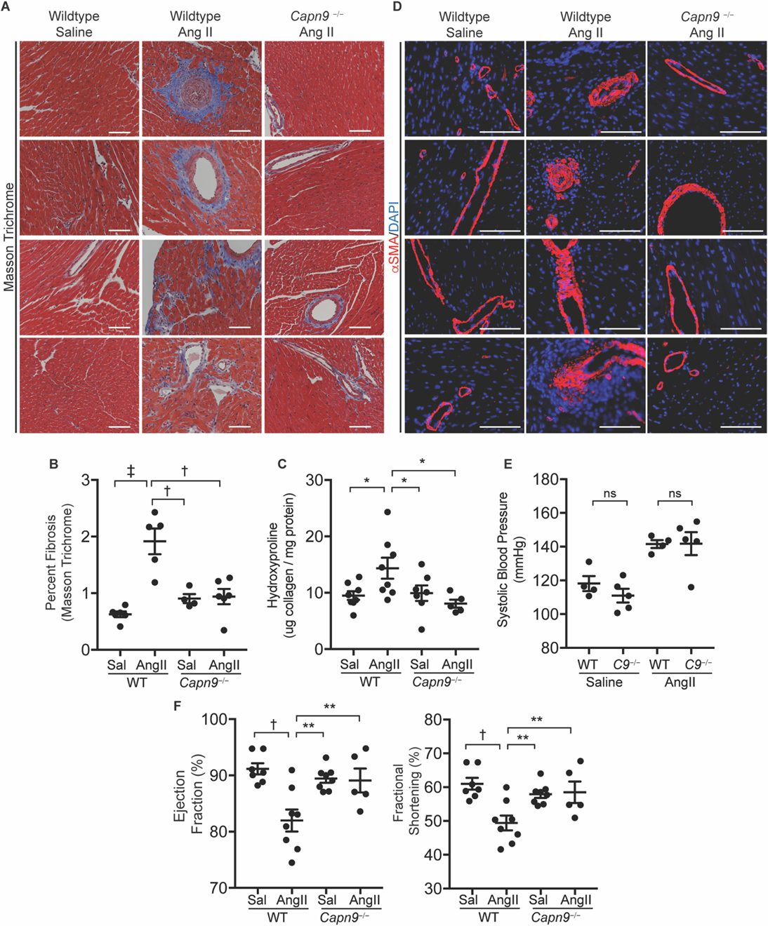
(A) Representative Masson trichrome stained slides of wildtype and Capn9-targeted mouse hearts from mice treated with saline or chronic Ang II infusion. Scale bar: 100 µm. (B) Quantification of blue stained collagen on trichrome slides by ImageJ (n = 4 to 6). (C) Collagen content in hearts from Capn9−/− or wildtype control mice treated with saline (Sal) or Ang II (n = 5 to 8). (D) Representative images of mouse hearts from groups as in (A), stained for SMA (red) or DAPI (blue). Scale bar: 100 µm. (E) Systolic blood pressure of wildtype and Capn9−/− (C9−/−) animals receiving Ang II infusion (n = 4 to 5). (F) Measures of left ventricular function including ejection fraction and fractional shortening in mice treated with Ang II (n = 5 to 8). Data are expressed as mean ± s.e.m. with statistical comparison between Ang II-treated wildtype mice and all other conditions. nsP > 0.05, *P < 0.05, **P < 0.01, †P < 0.005, ‡P < 0.001 by one-way ANOVA with Holm-Sidak post hoc test (B, D, F) and two-way ANOVA with Tukey’s post hoc test (E). Treatment effect was significant (P < 0.0001).