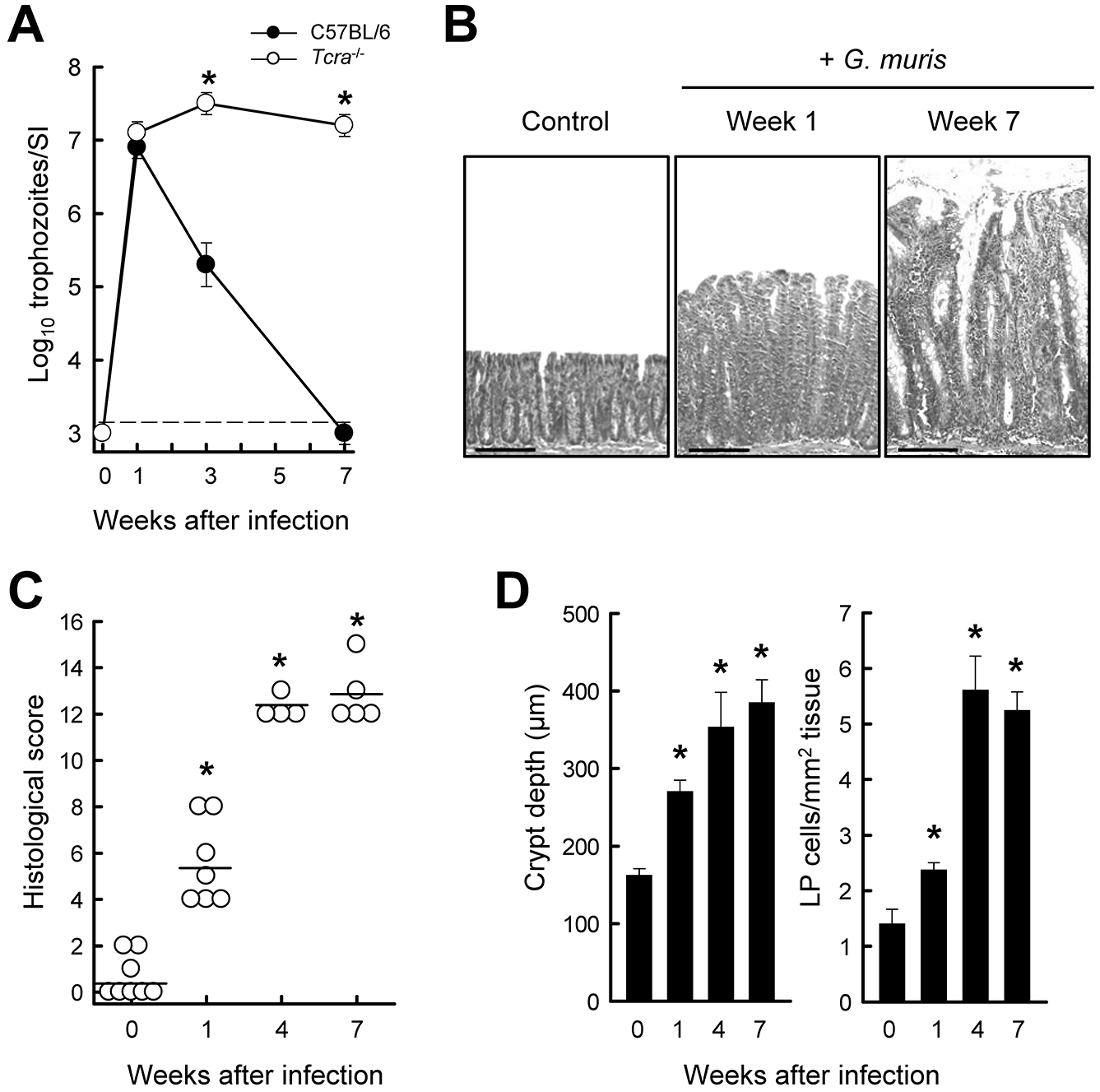
FIG. 2.

Susceptibility of Tcrα−/− mice to G. muris-induced colitis. Healthy age-matched (7- to 8-week-old) littermates of Tcrα−/− (open circles) and wild-type (solid circles) mice were infected with G. muris or left uninfected as controls, and followed for 7 weeks. (A) Trophozoite numbers in the small intestine were determined at the indicated times (mean ± SE; n≥3 mice/time point; *p<0.05 vs. wild-type at the same time). (B-D) Histological analysis of the colon in Tcrα−/− mice. Representative H&E-stained sections are shown in panel B (scale bars, 100 μm). Histological scores of colon damage and inflammation were determined (C; each data point represents one animal, horizontal lines represent mean values for each group; *p<0.05 vs. uninfected mice). Crypt depths and cell density in the lamina propria (LP) were quantified morphometrically (D; means ± SE, n≥4 mice/group; *p< 0.05 vs. uninfected mice).
