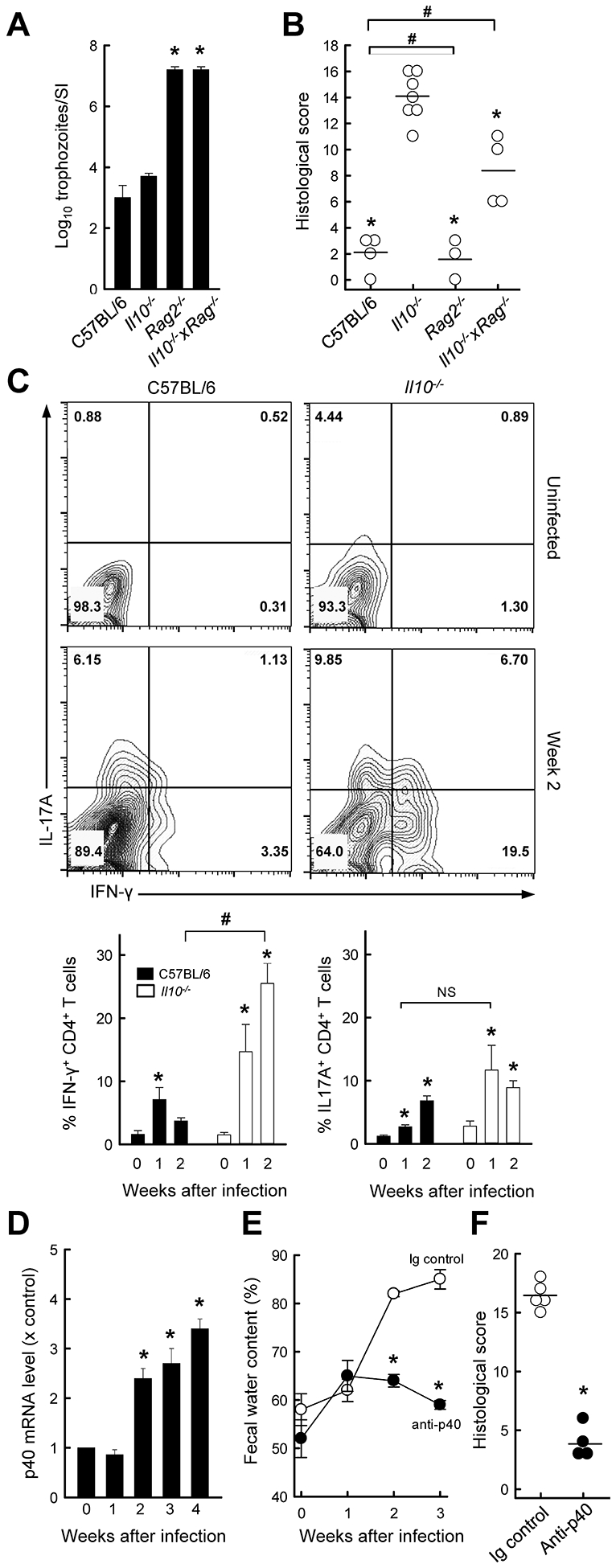
FIG. 3.

IL-12 p40 promotes colitis development after Giardia infection in IL-10−/− mice. (A, B) Il10−/−, Rag−/−, Il10−/− x Rag−/−, and C57BL/6 control mice were infected with G. muris. Three weeks after infection, parasites were enumerated in the small intestine (A), and histological analysis of the colon was done (B; each data point represent one animal, horizontal lines show mean values for each group; *p<0.05 vs IL-10−/− mice, #p<0.05 vs wild-type mice). (C) IL-10−/− mice (open bars) and wild-type C57BL/6 mice (solid bars) were infected with G. muris or left uninfected as controls (Week 0), and single cells were isolated from the colon lamina propria and analyzed by flow cytometry for IFN-γ and IL-17A expressing CD4+ T cells. Representative FACS contour plots are shown (top panel), and percentages of Th1 and Th17 cells are given (bottom panel; means ± SE, n≥7 mice/time; *p<0.05 vs. age-matched uninfected mice; #p<0.05 vs. wild-type mice at the same time; NS, not significant). (D) Quantitative RT-PCR analysis of IL-12 p40 mRNA in the colon of Il10−/− mice infected with G. muris (means ± SE, n≥7 mice/time; *p<0.05 vs. uninfected controls). (E, F) IL-10−/− mice were treated with anti-p40 or an isotype control IgG (Ig), and infected with G. muris. Fecal water content was assessed at the indicated times (E). Histological damage scores were determined three weeks after infection (F; each data point represents one animal, horizontal lines shown means for each group; *p<0.05 vs. Ig control).
