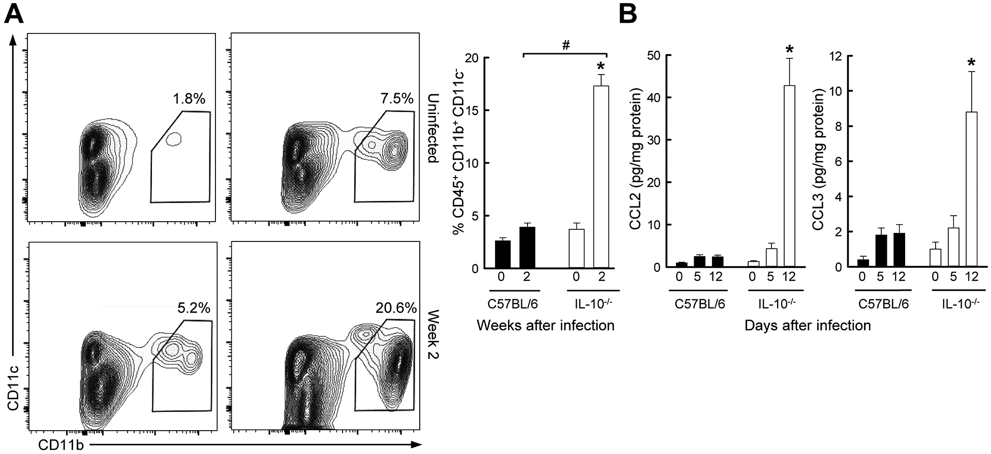
FIG. 5.

Colon infiltration with macrophages in Giardia-infected IL-10 deficient mice. Il10−/− mice (open bars) and wild-type C57BL/6 mice (solid bars) were infected with G. muris, or left uninfected (Week 0). (A) Single cells were isolated from the colon lamina propria and analyzed by flow cytometry for macrophages. Representative FACS contour plots are shown on the left, and percentages of macrophages are given on the right (mean ± SE, n≥6 mice/group; *p<0.05 vs. age-matched uninfected mice; #p<0.05 vs. wild-type mice at the same time). (B) Levels of CCL2 and CCL3 were determined in colon extracts by multiplex immunoassay (mean ± SE, n=5 mice/time; *p<0.05 vs. age-matched uninfected mice of the same genotype, #p<0.05 vs. wild-type mice at the same time).
