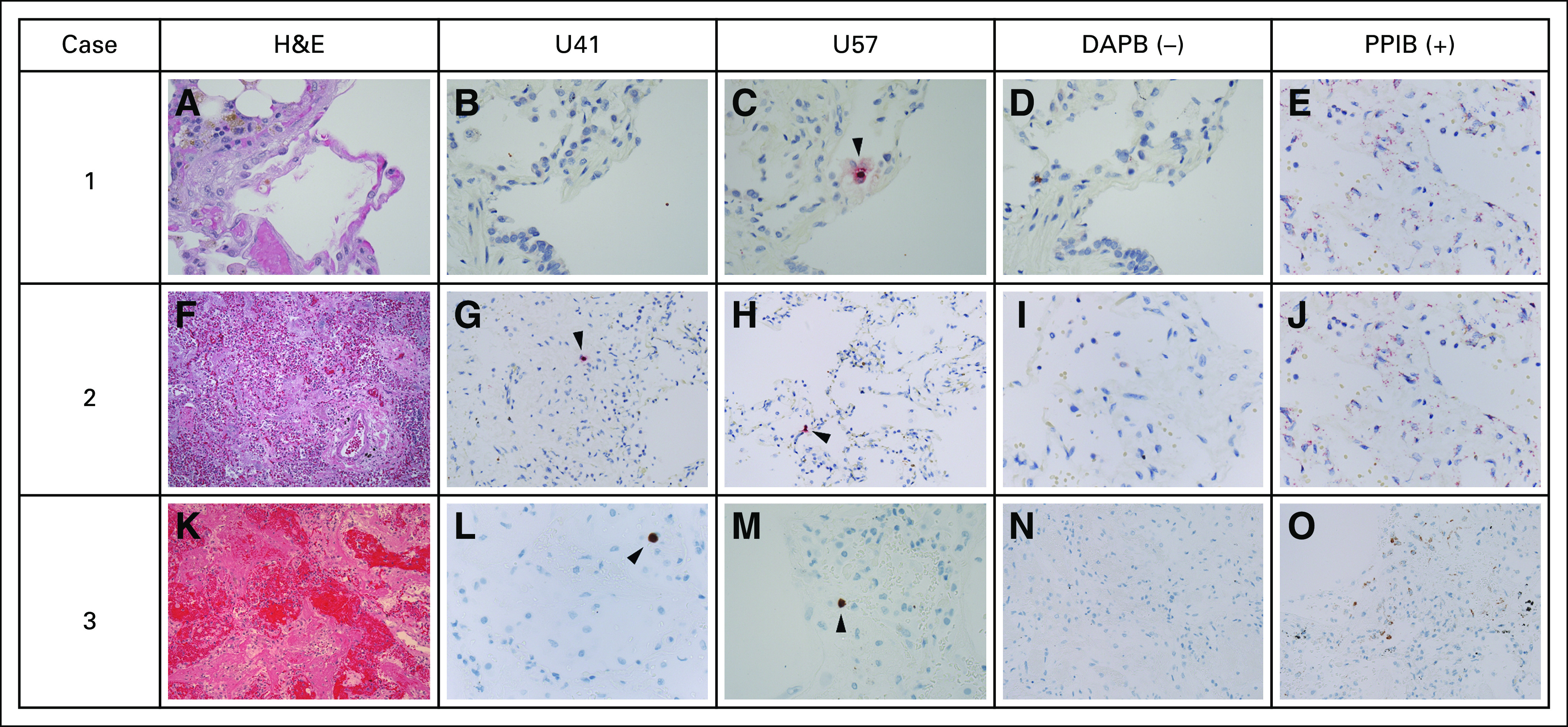
FIG 6.

RNAscope branched-chain RNA in situ hybridization (RISH) for HHV-6 messenger RNA transcripts (U41 and U57 transcripts) in formalin-fixed paraffin-embedded lung-tissue specimens from subjects with HHV-6B+ BALF. (A-O) The images under the columns labeled “U41” and “U57” correspond to HHV-6 RISH targeting HHV-6 early (U41) and late (U57) messenger RNA transcripts expressed during lytic viral infection. The images under the column labeled “DAPB” represent the negative control, which is a cocktail of probes targeting bacterial genes. The images under the column labeled “PPIB” represent the positive control, which is a cocktail of normally weakly expressed human control genes. Each row represents a subject with HHV-6B+ BALF sample collected within 10 days of the tissue-sample collection date. Only tissue samples that met prespecified quality of RNA preservation based on PPIB staining were included (Data Supplement). (A) In case 1, the subject was diagnosed with bacterial and fungal pneumonia. The hematoxylin and eosin (H&E) stain demonstrated a thin alveolar wall, some congested capillaries and hemosiderin pigment, but no overt viral cytopathic changes. (B) RISH (with red chromogen staining) for U41 was negative . (C) RISH for U57 demonstrated rare positive cells. In case 2, the subject was diagnosed with bacterial and fungal pneumonia. (F) The H&E stain demonstrated diffuse alveolar damage and organizing pneumonia with significant vascular congestion but no overt viral cytopathic changes. RISH (with red chromogen staining) for (G) U41 and (H) U57 demonstrated rare positive cells. In case 3, the subject was diagnosed with idiopathic pneumonia syndrome and diffuse alveolar hemorrhage. (K) The H&E stain demonstrated marked destruction of normal lung parenchyma with hemorrhage and diffuse alveolar damage. There was a comparatively mild inflammatory infiltrate without overt viral cytopathic changes. RISH (with brown chromogen staining) for (L) U41 and (M) U57 demonstrated rare positive cells. (C, G, H, L, M) The rare positive cells highlighted by the arrowheads are intraparenchymal, often seen within alveolar walls, and, therefore, favored to represent pneumocytes or lymphocytes. (D, I, N) The negative control stains for each case demonstrated minimal to no background staining. (E, J, O) The positive control stains for each case demonstrated good staining to indicate preservation of RNA in the samples.
