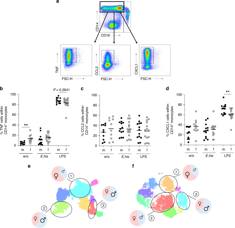
Fig. 4. TNF+, CXCL1+, and CCL2+ classical monocytes are more abundant in men.
a Gating strategy and representative dot plots showing cytokines produced by classical monocytes upon stimulation. PBMCs from men and women were stimulated for 6 h with E. histolytica lysate (0.1 mg/mL) or LPS (0.1 µg/mL), and intracellular expression of b TNF, c CCL2, and d CXCL1 by classical monocytes was measured by flow cytometry and compared to unstimulated controls (b nm/f w/o = 10/10; nm/f E. his = 13/14; nm/f LPS = 13/14; c nm/f w/o = 10/10; nm/f E. his = 13/14; and nm/f LPS = 13/14; d nm/f w/o = 10/10; nm/f E. his = 13/14; nm/f LPS = 13/14; pooled data from samples collected over a time frame of nine months; depicted are the means). Hierarchical Stochastic Neighbor Embedding (HSNE) analysis of e E. histolytica- CD14+CD16− and f LPS-stimulated CD14+CD16− classical monocytes. Cluster (1), TNF+CXCL1−CCL2−; cluster (2), TNF+CXCL1+CCL2−; and cluster (3), TNF+CXCL1+CCL2+ (calculated from classical CD14+ monocytes) (Table 2). P-values were calculated using two-tailed grouped analysis: Mann–Whitney test (b, d), **P < 0.01. Source data are provided as a Source Data file.

