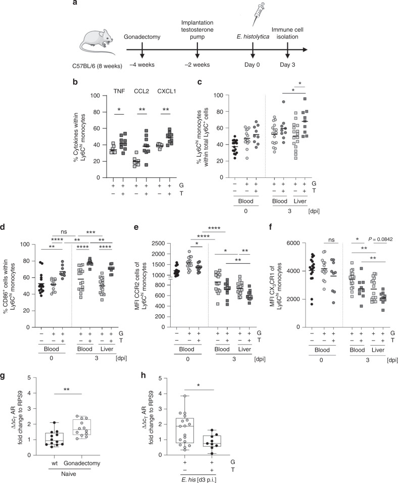
Fig. 7. Testosterone modulates classical monocytes towards a proinflammatory phenotype.
a Mice were either gonadectomized (G) or gonadectomized (4 weeks before infection, 8 weeks of age) and substituted with testosterone (T) (2 weeks before infection, 10 weeks of age). b Intracellular expression of TNF, CCL2, and CXCL1 by liver-derived Ly6Chi monocytes from G and T male mice 3 days following intrahepatic E. histolytica infection, as analyzed by flow cytometry. (TNF: nG/T = 7/9; CCL2: nG/T = 7/9, CXCL1: nG/T = 7/9; one experiment; depicted are the means). c Percentage of Ly6Chi monocytes in the blood and liver of naïve or E. histolytica-infected G and T male mice (blood: nwt/G/T d0 = 16/14/9; nG/T d3 = 16/9; liver: nG/T d3 = 16/9; one experiment; depicted are the means). Percentage of d CD86+ cells and e MFI of CCR2 and f CX3CR1 Ly6Chi monocytes in the blood and liver of naïve or E. histolytica-infected G and T male mice (d blood: nwt/G/T d0 = 16/14/9; nG/T d3 = 16/9; liver: nG/T d3 = 16/9; e blood: nwt/G/T d0 = 16/14/9; nG/T d3 = 16/9; liver: nG/T d3 = 16/9; f blood: nwt/G/T d0 = 16/14/9; nG/T d3 = 16/9; liver: nG/T d3 = 16/9; one experiment; depicted are the means). g qPCR analysis of androgen receptor (AR) expression of RNA isolated from PBMCs of naïve sham operated or gonadectomized mice (nsham/G = 12; one experiment; depicted are box ranging from 25th to 75th percentile and whiskers from min to max including the median). h qPCR analysis of androgen receptor (AR) expression of RNA isolated from PBMCs of E. histolytica-infected G and T mice (nG/T = 17/9; pooled data from two independent experiments; depicted are box ranging from 25th to 75th percentile and whiskers from min to max including the median). P-values were calculated using two-tailed grouped analysis: Student’s t-test (b) or Mann–Whitney test (c, d, e–h), *P < 0.05; **P < 0.01; ***P < 0.001; ****P < 0.0001. Source data are provided as a Source Data file.

