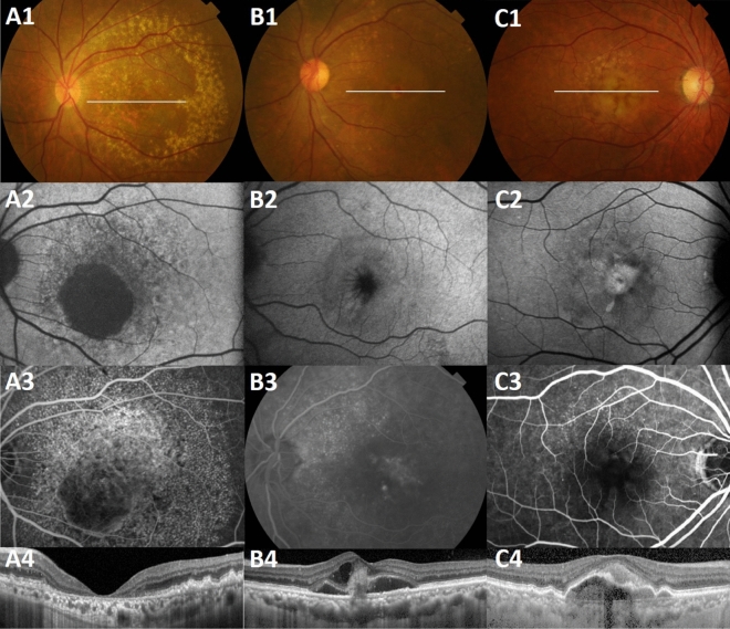
Figure 2.
Macular complications of cuticular drusen. (A) Geographic atrophy is seen on (A1) CFP, (A2) FAF, (A3) FA, and (A4) OCT in a 65-year-old man with cuticular drusen. Geographic atrophy appears in both eyes symmetrically. Large drusen are also visible on CFP and OCT. (B) Type 3 choroidal neovascularization is seen on (B1) CFP, (B2) FAF, (B3) FA, and (B4) OCT in a 67-year-old woman with cuticular drusen. (C) Drusenoid pigment epithelial detachment and acquired vitelliform lesion appear as (C1) round yellow lesions on CFP, (C2) intense hyperautofluorescence on FAF, (C3) hypofluorescence in the early phase of FA, and a (C4) subretinal location of vitelliform material with pigment epithelial detachment is seen on OCT, in a 74-year-old man. The position of the OCT line scans is shown in the CFP as a white transverse line. CFP color fundus photograph, FA fluorescein angiography, FAF fundus autofluorescence, OCT optical coherence tomography.

