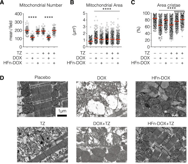
Figure 2.
Analysis of HFn-DOX and TZ mitochondrial cardiotoxicity. Hearts from treated mice were fixed with glutaraldehyde and embedded in epoxy-resin. At least 10 TEM images of ultrathin heart sections have been acquired at 4,200 and 11,500 magnifications for each experimental group. Quantification of mitochondria area and area occupied by cristae were performed on at least 10 images/group, measuring at least 100 mitochondria/sample, while count of mitochondria number were performed on at least 10 images/group. Values represent the mean mitochondria number ± s.e. (A), the mean mitochondrial area ± s.e. (B) and the percentage of mitochondrial area occupied by cristae ± s.e. (C). Statistical significance vs. Placebo ****P < 0.0001; vs TZ ####P < 0.0001 (Kruskal-Wallis test). (D) Representative images of mitochondria from hearts excised at day 24 (n = 3/group) from treated mice acquired at 11,500 magnifications evidenced mitochondria morphological alterations.

