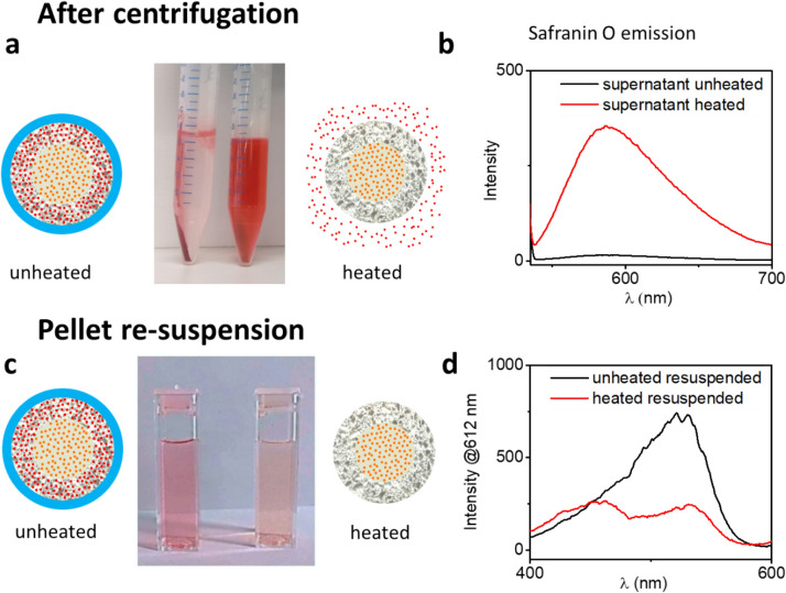
Figure 3.
(a) A picture of centrifugation tubes containing the pellets and supernatants of solutions of unheated (left) and heated (74 °C, right) solutions of core–shell–hull (dotriacontane) nanoparticles after centrifugation, with the corresponding schematic representations of the state of the nanoparticles. (b) The fluorescence emission signal of the supernatant obtained from the centrifugation tubes, upon excitation at 530 nm. (c) An image of cuvettes containing solutions of the re-suspended nanoparticles taken from the pellets in the centrifugation tubes. (d) Fluorescence spectroscopy of the solutions displayed in (c), conducted by sweeping the excitation wavelength and measuring the resulting emission at the 612 nm wavelength.

