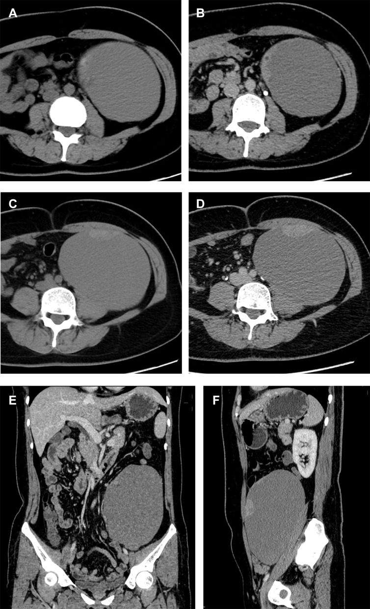
Figure 2.
Pre-operative imaging of a 44-year-old female with intraepithelial carcinoma in bPRMC (Case 10). Unenhanced and enhanced axial CT scan (A and B) reveals an oval-shaped, well-circumscribed cyst with a thick irregular wall and contrast enhancement in the venous phase, while axial CT scan (C and D) shows a broad-based mural nodule on the front wall with contrast enhancement in the venous phase. CT scan in the coronal (E) and sagittal (F) planes also demonstrates an irregular cyst wall and mural nodules in the venous phase.

