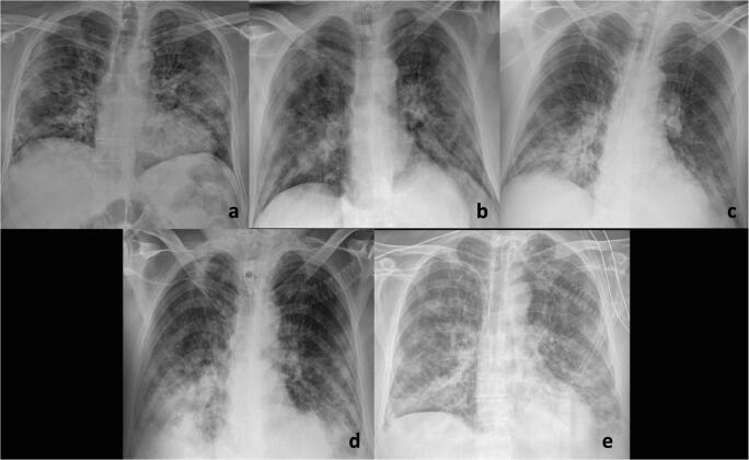
Fig. 2.
A 72-year-old man with fever and cough for 7 days and COVID-19 pneumonia in chest imaging. Serial chest radiographs performed at admission (a), 12 h after tracheal intubation in ICU (b), 5 days (c), 10 days (d), and 12-days (e) of ICU demonstrate progressive worsening of the bilateral lung opacities and consolidations

