Figure 3.

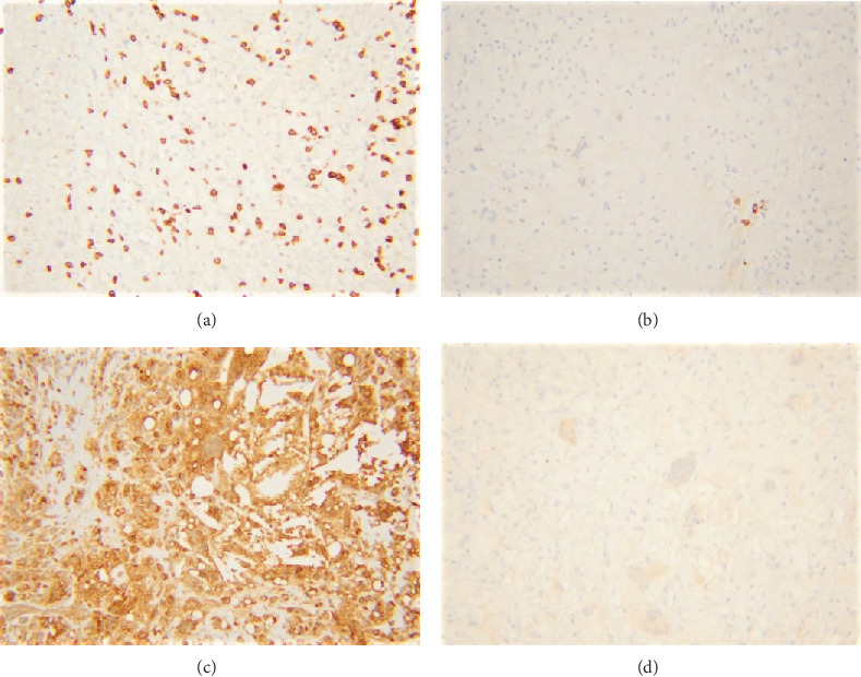
Immunohistochemical stains reveal scattered CD3-positive T-cells (a), rare CD20-positive B-cells (b), and majority of the cells staining positive for CD68 (c). Pancytokeratin stain is negative (d).

Immunohistochemical stains reveal scattered CD3-positive T-cells (a), rare CD20-positive B-cells (b), and majority of the cells staining positive for CD68 (c). Pancytokeratin stain is negative (d).