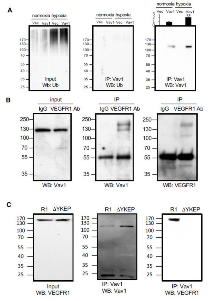
Figure 2.
Vav1 binds to vascular endothelial growth factor receptor 1 (VEGFR1). (A) Either Flag-tagged Vav1 or empty vector control were transduced into HUVEC cells and incubated under normoxic or hypoxic conditions. Cell lysates were subjected to Vav1 immunoprecipitation and subsequent Western blot analysis for ubiquitin (middle panel) or Vav1 as a control (right panel). Ubiquitin levels were also detected in both control and Vav1 cells exposed to hypoxia from the input of the immunoprecipitation (left panel). (B) VEGFR1 and Vav1 were immunoprecipitated by anti-VEGFR1 antibody or control IgG from the cell lysate of HUVEC cells. Vav1 was probed from the input lysate (left) and pulldown product (center). VEGFR1 was detected from the pulldown product (right). (C) Overexpressed wild type (R1) or YKEP motif deleted (ΔYKEP) VEGFR1 were co-IPed with Vav1 antibody. The input was probed with VEGFR1 antibody (left). The pulldown product was probed against Vav1 (center) or VEGFR1 antibody (right). Mean ± SD, * p < 0.05, ** p < 0.01

