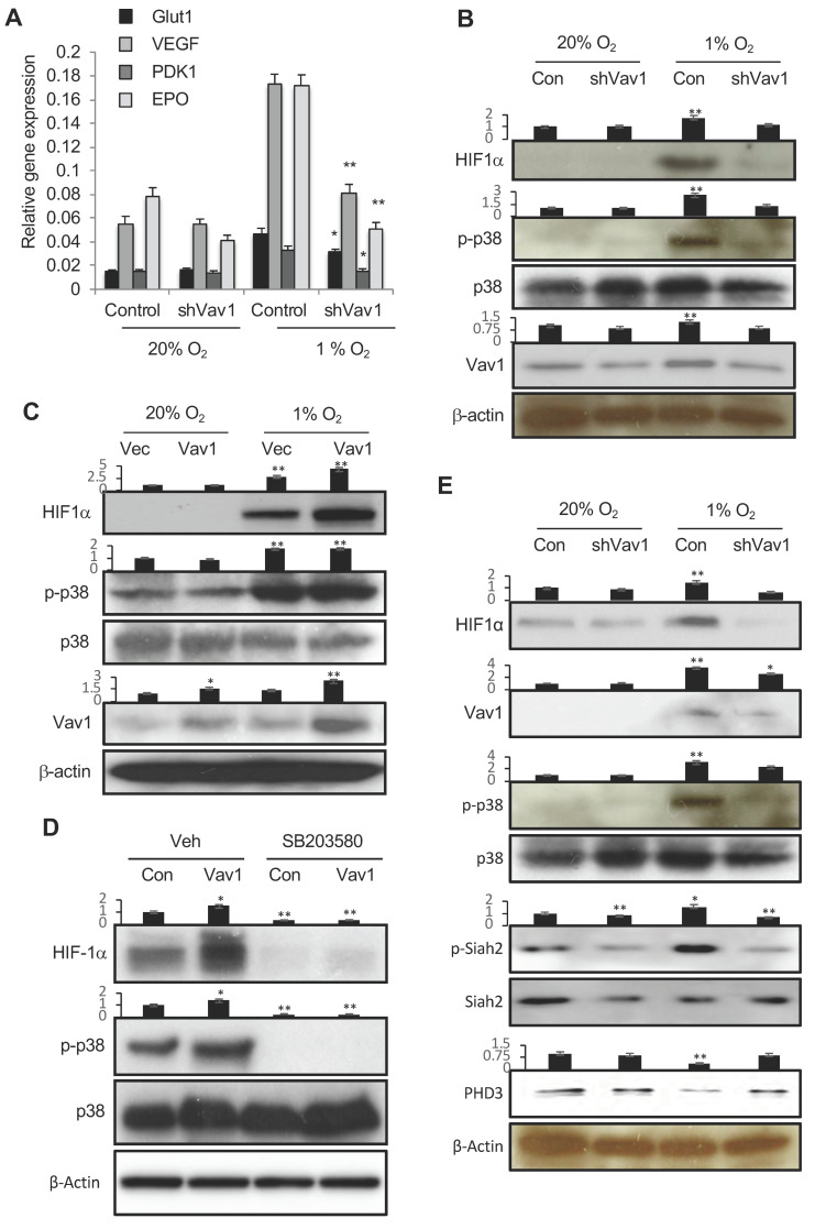
Figure 4.
Vav1 regulates HIF1α via p38 MAPK. (A) HUVECs were transfected with a control vector or shVav1 vector for 24 h, followed by incubation either in normoxia or hypoxia for another 24 h. HIF-1 target gene expression was analyzed by qRT-PCR. * p < 0.05 and ** p < 0.01 compared to corresponding control transfected cells in hypoxia (mean ± SD). Each experiment was performed in triplicate and repeated three times. (B) Control vector- or shVav1 vector-transfected HUVECs were incubated under normoxia or hypoxia conditions for 24 h. The levels of phosphor-p38, HIF-1α, p38 and Vav1 were analyzed by Western blot. (C) Identical procedures and measurements as in (B) except the cells were transfected with a control vector or Vav1 expression vector. (D) Control or Vav1 expression vector-transfected HUVECs were incubated in the absence or presence of SB203580 at 10 μM in hypoxia for 24 h. The levels of HIF-1α, p38 and phospho-p38 were analyzed by Western blot. (E) Control vector- or shVav1 vector-transfected HUVECs were incubated under normoxic or hypoxic conditions for 24 h. The levels of phospho HIF-1α, Vav1, P-p38, p38, P-siah2, Siah2 and PHD3 were analyzed by Western blot. Mean ± SD, * p < 0.05, ** p < 0.01. The whole western blot images please find in Figure S3.

