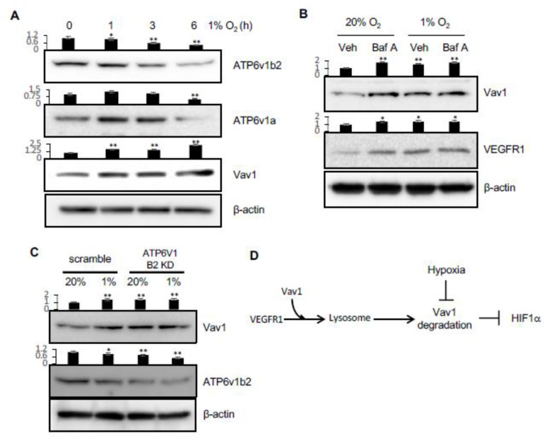
Figure 5.
Hypoxia regulates lysosomal activity via v-ATPase and Vav1 levels. (A) HUVEC cells were incubated in hypoxia for 0, 1, 3 or 6 hours. Vav1, ATP6v1a, and ATP6v1b2 were measured in the total lysates. (B) HUVEC were incubated in normoxia or hypoxia for 5 h in the presence or absence of 100 nM of Bafilomycin A (Baf A). Vav1 and VEGFR1 were measured by Western blot of the total lysates. (C) HUVEC were transduced with ATP6v1b2 shRNA-expressing lentivirus or scrambled shRNA virus for 72 h, and then incubated in normoxia or hypoxia for 5 h. Vav1 and ATP6v1b2 levels were measured by Western blot of the total lysate. (D) VEGFR1 carries Vav1 to the lysosome in the cell. Vav1 is degraded in the lysosome, which downregulates HIF1α. Hypoxia blocks the lysosomal degradation. Mean ± SD, * p < 0.05, ** p < 0.01. The whole western blot images please find in Figure S4.

