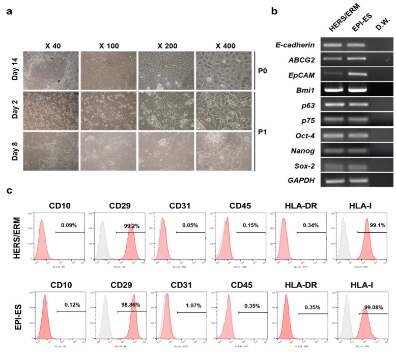
Figure 2.
Differentiation of human embryonic stem cells (hESC) into dental epithelial-like stem cells (EPI-ES). (a) Morphological change during hESC differentiation into EPI-ES. After 14 days on the feeder layer, typical epithelial cell-like cuboidal- or polygonal-shaped appearances were observed around the hESC clumps. During two to eight days after subculture, cells with these morphologies underwent colony-forming proliferation. (b) Gene expression of EPI-ES after 14 days of induction. All samples, as well as HERS/ERM, were positive for epithelial stem cell markers and stemness markers. (c) Expression of surface antigens of EPI-ES. Both primary HERS/ERM and all epithelial-like cell lines were positive for mesenchymal markers (CD29) and HLA type I, but negative for hematopoietic cell markers (CD10, CD45, and HLA-DR) and an endothelial cell marker (CD31). All data were replicated three times.

