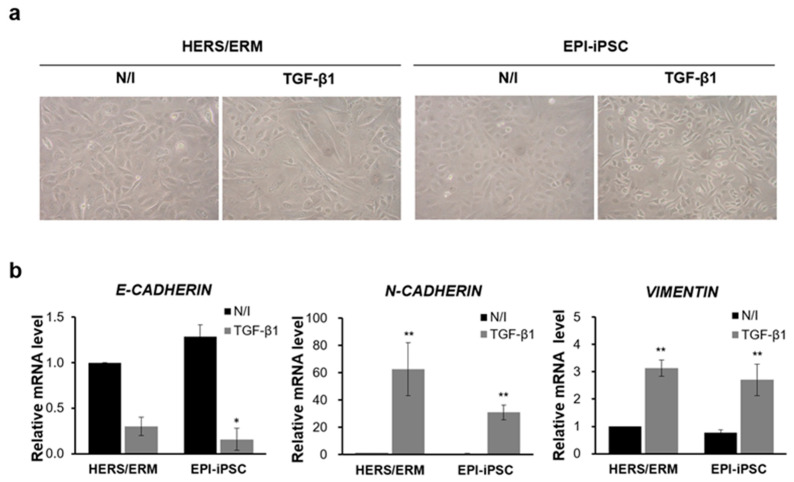
Figure 5.
Epithelial-mesenchymal transition (EMT) of HERS/ERM cells and the EPI-iPSC cell line. The EMT was induced by TGF-β1 for 48 h. (a) Morphology of the EPI-iPSC cell line after 48 h of TGF-β1 treatment. All of these cells lost epithelial cell polarity and cell-to-cell contact. (b) EMT-related gene expression of the EPI-iPSC cell line after EMT induction. When all cell types were treated with TGF-β1, the gene expression of N-cadherin and Vimentin was increased in primary HERS/ERM and epithelial-like cells. However, the levels of E-cadherin were decreased. All data shown are the mean ± S.D. from the levels of three replicates. Data are presented as the mean ± SD, n = 6 per group. ** p < 0.01, * p < 0.05. N/I: no induction.

