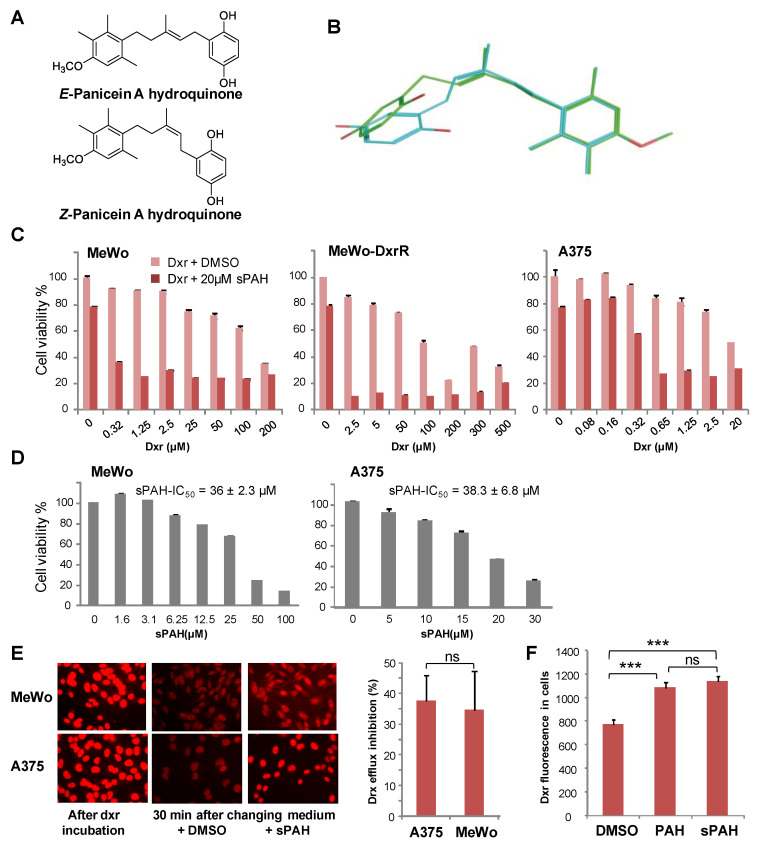
Figure 2.
The chemically synthesized PAH is as effective as natural PAH in increasing cytotoxicity and inhibiting the efflux of doxorubicin. (A) Stereoisomers of PAH. (B) 3D structures of E and Z forms of PAH are very similar. The E form is in green and the Z form is in cyan. (C) sPAH increases doxorubicin cytotoxicity in various melanoma cell lines. Cell viability was measured after treatment with increasing concentrations of dxr in the presence of DMSO or 20 µM sPAH on MeWo cells, MeWo cells rendered resistant to dxr (MeWo-DxrR) and A375 cells. (D) sPAH cytotoxicity against melanoma cells. Cell viability was measured after treatment with increasing concentrations of sPAH on A375 or MeWo cells. The mean sPAH-IC50 values calculated from 3 independent experiments are represented. (E) sPAH inhibits the dxr efflux activity of Ptch1. A375 or MeWo cells were seeded on coverslips and incubated with dxr. After 2 h, 3 coverslips were fixed for dxr loading control. The other coverslips were incubated with DMSO or sPAH and fixed. Dxr fluorescence was imaged and quantified using ImageJ software for about 100 cells per condition per experiment. The inhibition of dxr efflux by sPAH for each cell lines is reported. (F) Synthetic PAH inhibits the dxr efflux activity of Ptch1 as efficiently as natural PAH. Dxr fluorescence in MeWo cells was quantified after 30 mins in buffer containing DMSO, natural PAH, or synthetic PAH as described in E. All histograms represent the mean ± SEM of 3 independent experiments. Significance is attained at p < 0.05 (*) (***: p < 0.0005), ns: no significant difference.

