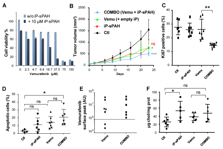
Figure 8.
sPAH enhances the effect of vemurafenib on BRAFV600E melanoma cells xenografted in mouse. (A) sPAH encapsulated in i-Particles increases the cytotoxicity of vemurafenib. Cell viability was measured after treatment with increasing concentrations of vemurafenib with or without iP-sPAH in A375 cells. (B) The addition of iP-sPAH to vemurafenib significantly inhibits the growth of tumors. Mice were injected subcutaneously with A375 cells and treated with either empty i-Particles (empty iP) (Ctl), vemurafenib and empty iP, iP-sPAH, or a combination of vemurafenib and iP-sPAH. Tumor size was measured every 3 to 4 days. At day 23 animals were sacrificed, and tumors were excised and subsequently snap frozen using OCT. (C) The addition of iP-sPAH to vemurafenib significantly reduced the number of proliferative cells in tumors. Tumor sections were submitted to Ki67 immunostaining for quantification of proliferative cells using fluorescence microscopy and ImageJ software. (D) The addition of iP-sPAH to vemurafenib significantly increases the number of apoptotic cells in tumors. Tumor sections were submitted to the colorimetric DeadEND TUNEL System to determine the number of apoptotic tumor cells in each tumor. (E) The addition of iP-sPAH to vemurafenib increases the amount of vemurafenib in tumors. The amount of vemurafenib in each tumor extract was quantified by mass spectrometry. (F) iP-sPAH increases the amount of cholesterol in tumors. Sterols were extracted from tumor homogenates and analyzed by GC–MS. All data presented are the mean ± SEM of 3 independent experiments. Significance is attained at p < 0.05 (*) (**: p < 0.005, ***: p < 0.0005); ns: no significant difference.

