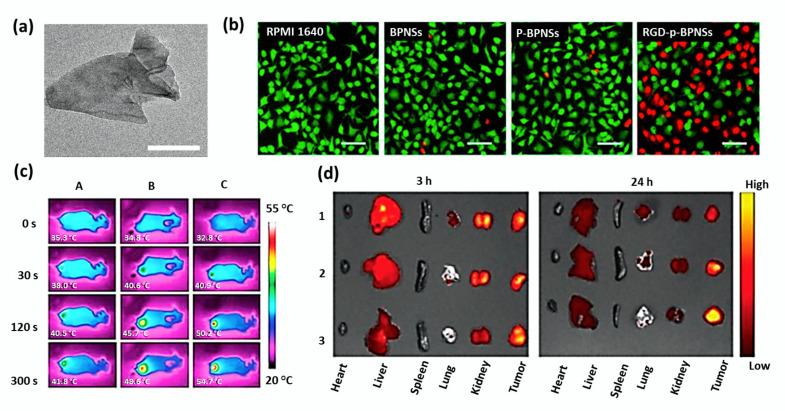
Figure 6.
(a) TEM image of bare BPNSs with scale bar 50 nm. (b) Inverted fluorescence microscope images of A549 cells (scale bar 100 μm) treated with RPMI 1640 cell culture medium, bare BPNSs, p-BPNSs (1-pyrenebutyric acid-modified BPNSs), and RGD-p-BPNSs after 30 min of incubation followed by NIR irradiation laser (808 nm, 1.0 W/cm2, 10 min). Propidium iodide (red, dead cells) and Calcein-AM (green, live cells) were used for staining the cells. Adapted with permission from [104]. (c) In vivo IR thermal images of mice treated with A: PBS; B: BP@PDA-PEG; C: BP@PDA-PEG-Apt. (d) Ex vivo fluorescence images of tumors and major organs captured at 3 and 24 h of post-administration of free DOX (1), BP-R-D@PDA-PEG (2), and BP-R-D@PDA-PEG-Apt (3). Adapted with permission from [54].

