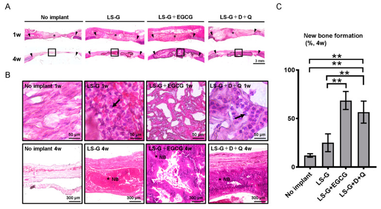
Figure 3.
Histological evaluation of bone defects. Low (A) and high (B) magnification microscopic images of bone defects stained with hematoxylin–eosin one or four weeks after surgery. The image in (B) represents magnified squares in (A). Black triangles, edge of created bone defects. Black arrows, leucocytes. Black broken lines in (A) show newly formed bone. * NB, new bone; D, dasatinib; Q, quercetin. (C) Histomorphometric analysis of newly formed bones using the samples of H-E staining. Data are expressed as mean ± SD. ** p < 0.01. LS-G, implantation of LPS sustained-release gelatin sponges in bone defects; LS-G+EGCG, implantation of LS-G chemically modified with EGCG in bone defects; LS-G+D+Q, oral administration of senolytics (D+Q) with implantation of LS-G in bone defects.

