Figure 2.
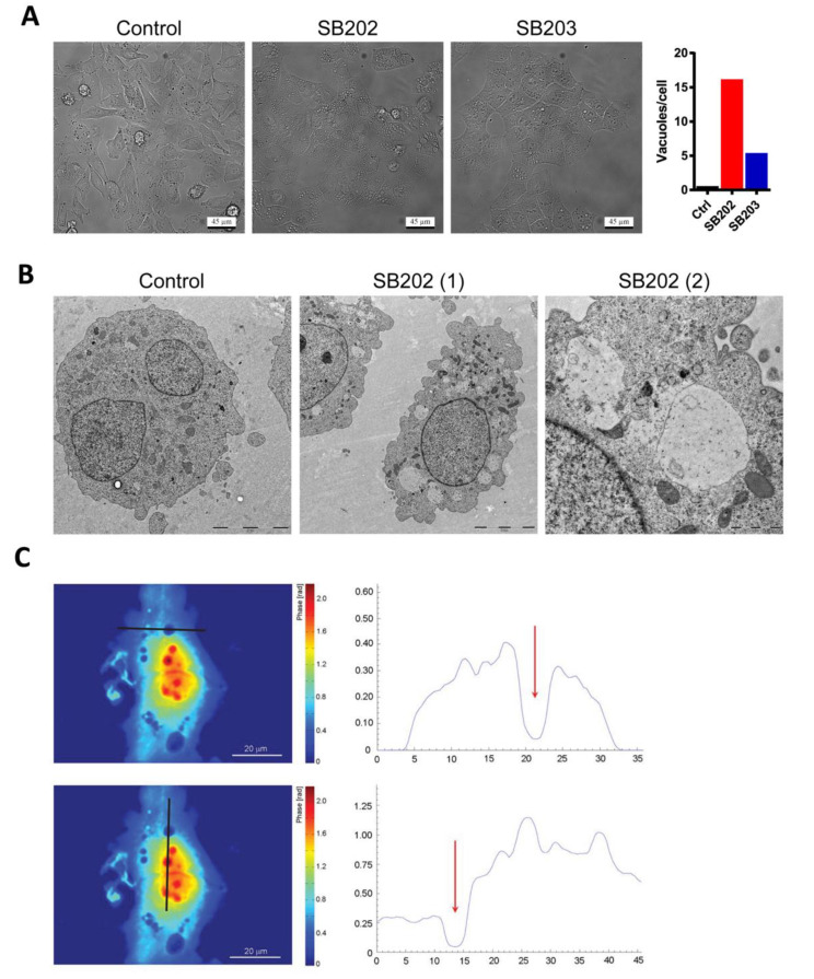
Pyridinyl imidazole compounds induce vacuolization of cytoplasm in BRAF-mutated human melanoma cells. A375 cells were treated with DMSO (control), SB202190 (SB202; 10 μM), and SB203580 (SB203; 10 μM). (A) Phase-contrast light microscopy of living cells after 12 h treatment with pyridinyl imidazole p38 MAPK inhibitors. Scale bar: 45 µm. The representative graph shows the mean number of vacuoles per cell quantified using ImageJ/Fiji (find maxima—bright spots above a certain threshold). Dying rounded cells were excluded from the analysis. Similar results were obtained in three independent experiments. (B) The content of vacuole-like vesicles was visualized by electron microscopy 24 h post-treatment with SB202190 (SB202). Control and SB202(1)—scale bar 5 µm. SB202(2)—scale bar 1 µm. Two independent experiments showed similar results. (C) Quantitative phase-imaging analysis of cellular dry mass in melanoma cells treated for 12 h with SB202190. The zero level of dry mass density was defined as the density of the observation medium. The areal density distribution of the dry mass was quantified in profiles (right panels) indicated by dark lines. Red arrows highlight the position of vacuoles. Two independent digital holography microscopy experiments showed similar results.

