Figure 2.
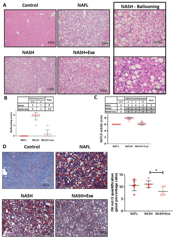
Effect of exercise on liver histology in mice fed a choline-deficient high-fat diet (CD-HFD). (A) Microscopy of hematoxylin and eosin (H&E)-stained liver sections showing diffuse macrovesicular steatosis in the NAFL, NASH and NASH + exercise (EXE) groups and the presence of ballooned hepatocytes only in the NASH sedentary group ). (B) Frequency table and dot plot comparing the ballooning score in the NASH sedentary and NASH+EXE groups. Ballooning was significantly lower in the NASH + EXE group (Fisher’s exact test, p = 0.005). (C) Frequency table and dot plot showing the NAFLD activity score in the NAFL, NASH sedentary, and NASH + EXE groups. The score was significantly lower in NASH + EXE than in the NASH sedentary group (Fisher’s exact test with Freeman–Halton extension, NASH vs. NASH + EXE, p < 0.0001). (D) Oil Red O staining comparing neutral lipid content of control, NAFL, NASH sedentary, and NASH + EXE livers. The quantification of lipid staining (right panel) was done with MetaMorph® analysis software. Lipid content was lower in the NASH + EXE livers (unpaired t-test; * p < 0.05).

