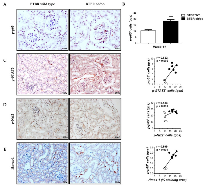
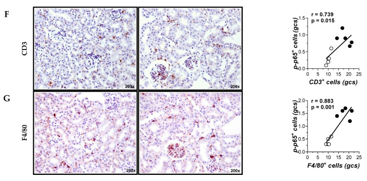
Figure 3.
Correlation between glomerular Nuclear factor-κB (NF-κB) activation and immunohistochemical kidney markers related to inflammation. Representative images (magnification 400× and 200×) of immunodetection of p-p65 (A), p-STAT3 (B), p-Nrf2 (C), Hmo×-1 (D), CD3 T cells (F) and F4/80 macrophages (G) in kidney sections; (B) Quantification of p-p65 positive cells in glomerular cross sections (gcs). Data shown as mean ± SD of n = 5–6 mice/group. **** p < 0.001 vs. BTBR wild type (WT) mice; (C–G) Pearson’s correlation analysis of glomerular p-p65 with different histological markers. White symbols indicate BTBR wild type; black symbols, BTBR ob/ob.


