Figure 4.
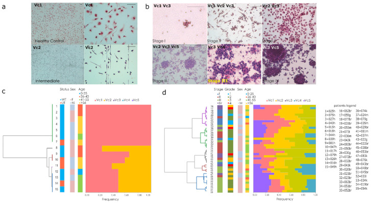
Cytology landscape in Blood-derived cultures (a) representative BDCs with lympho-monocytes elements (Vc1) and endothelial cells (Vc2) of control (Scale bars 100 μm). (b) Representative BDCs of cancer patients with atypical elements (Vc3), endothelial cells (Vc2), cluster and mitotic figures (Vc4, Vc5), i.e., in stage I a thyroid tumor case; stage II breast lobular (Vc1, Vc2, Vc3) and ductal (Vc2, Vc3) cases; stage III melanoma (Vc2, Vc3, Vc5) and NSCLC (Vc3, Vc4) cases; stage IV colon adenocarcinoma (scale bars 100 μm). (c,d) Hierarchical clustering based on Ward’s method for healthy (c) and cancer patient (d) each slides.

