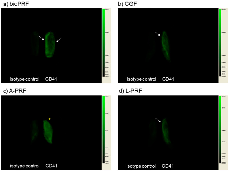
Figure 1.
NIR images of compressed half PRF matrices: (a) bio-PRF (horizontal, fast spin); (b) Concentrated growth factors (CGF) (fixed angle, fast programmed spin); (c) A-PRF (fixed angle, slow spin); (d) L-PRF (fixed angle, fast spin). White arrows represent platelet localization. An asterisk denotes homogeneous platelet distribution.

