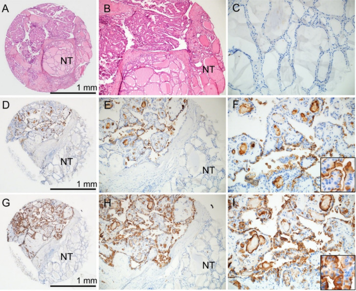
Figure 1.
Immunohistochemical staining for CD10 and CD15 on the tissue microarray of papillary thyroid carcinoma. (A) The tissue core contains tumor and non-tumor (NT) areas (hematoxylin and eosin stain). (B) The higher power view of the tissue core shows papillary thyroid carcinoma and NT adjacent to tumor. (C) NT in the same case is negative for both CD10 and CD15. Tumor cells in the same case are positive for both CD10 (D–F) and CD15 (G–I) immunostaining. (F) Inset shows CD10 staining at the apical surface of tumor cells. (I) Inset shows CD15 cytoplasmic and membranous staining in tumor cells; ×40 (A,D,G), ×100 (B,E,H), ×200 (C,F,I), inset (×400).

