Figure 1.
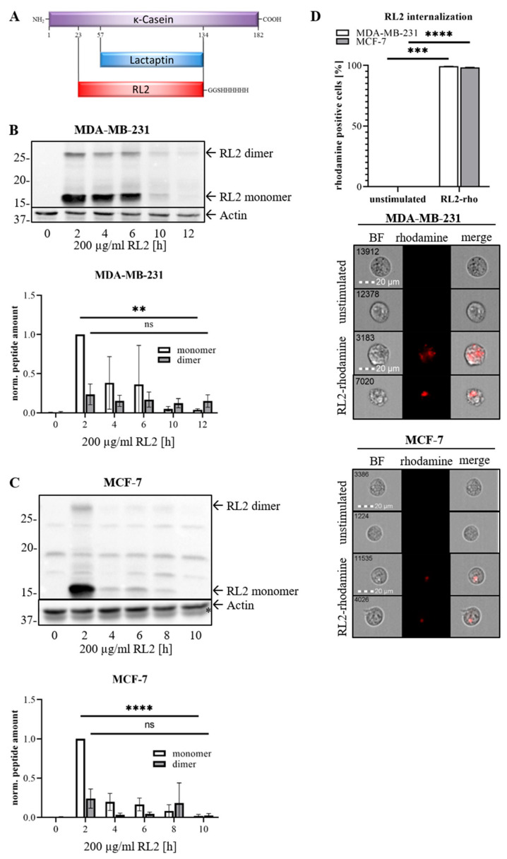
MDA-MB-231 and MCF-7 cells internalize RL2 and degrade it in a time dependent manner. (A) Scheme of RL2, Lactaptin and κ-Casein proteins. (B) MDA-MB-231 and (C) MCF-7 cells were treated with 200 µg/mL RL2 for indicated time intervals. Extracellular RL2 was removed by washing during cell harvest. RL2 was detected in the cellular lysates by Western Blot using anti-κ-Casein antibody. One representative Western Blot out of three independent experiments is shown. Quantification was done for three independent experiments. The statistical analysis was performed by one-way ANOVA test. (D) MDA-MB-231 and MCF-7 cells were treated with 50 µg/mL Rhodamine-labelled RL2 for four hours and analyzed with FlowSight® for RL2 internalization. Bottom: At the left column single cells are shown in bright field (BF) channel, at the right column merging of Rhodamine signal and bright field image is performed for representative single cells. Top: The amount of Rhodamine-RL2 positive cells are presented from three independent experiments. The statistical analysis was performed by paired student t-test. Relative Western Blot quantifications of Figure 1B,C are shown in Figure S1. ns (not significant; p > 0.05), ** (significant; p < 0.01), *** (significant; p < 0.005), **** (significant; p < 0.001).

