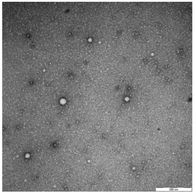
Figure 1.
Representative electron microscopy picture by negative staining showing their typical appearance and size range. The exosomes were isolated as described previously from the plasma of a head and neck squamous cell carcinoma (HNSCC) patient [11].

