Figure 1.
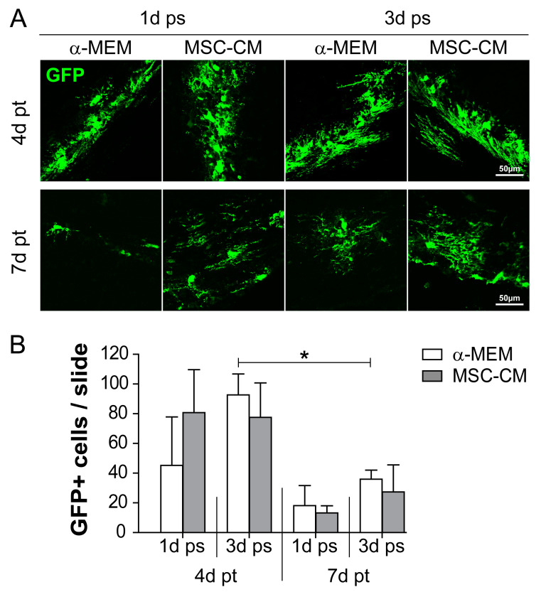
Survival rate of green fluorescent protein (GFP)-expressing cells after transplantation into the mouse brain. (A) Immunohistochemical staining was used to identify transplanted GFP-positive adult neural stem cells (aNSCs) (green) at 4 and 7 days post-transplantation (pt) into the mouse brain. aNSCs were pre-stimulated (ps) for either 1 or 3 days (1d ps and 3d ps, respectively) with α-minimum essential medium (α-MEM) or mesenchymal stem cell (MSC)-conditioned medium (MSC-CM). Representative pictures were taken from white (corpus callosum) / grey (cortex) matter implantation sites. (B) Quantitative evaluation of the total number of GFP-positive cells per slide including both grey and white matter. Statistical significance was calculated using a two-way ANOVA with Bonferroni posttest: * p ≤ 0.05. Data were generated on the basis of the following animal numbers (n): 1d ps and 4d pt, α-MEM and MSC-CM n = 4; 3d ps and 4d pt, α-MEM n = 5 and MSC-CM n = 4; 1d ps and 7d pt, α-MEM and MSC-CM n = 3; 3d ps and 7d pt, α-MEM and MSC-CM n = 4.

