Figure 1.
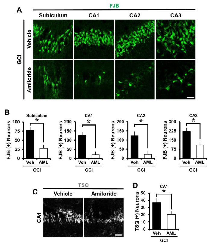
Amiloride treatment decreased the number of degenerating neurons and zinc accumulation after global cerebral ischemia (GCI). GCI-induced hippocampus neuronal death was confirmed in the subiculum (Sub), cornus ammonis 1 (CA1), CA2, and CA3 regions after ischemic insult. Zinc accumulation was confirmed in the CA1 region after ischemic insult. (A) Fluorescent images show degenerated neurons in the Sub, CA1, CA2, and CA3 regions. Intraperitoneal post-treatment with amiloride (10 mg/kg) reduced neuronal death in the Sub, CA1, CA2, and CA3 regions at 24 h after ischemia. Scale bar = 20 μm. (B) Bar graph displaying the quantification of degenerating neurons in the hippocampal regions. The number of FJB (+) neurons was decreased in the amiloride-injected (10 mg/kg) group in the Sub, CA1, CA2, and CA3 regions compared with the vehicle-treated group (GCI-vehicle, n = 8; GCI-amiloride, n = 8). (C) Representative images show N-(6-methoxy-8-quinolyl)-para-toluenesulfonamide (TSQ) (+) neurons in the CA1 region. Scale bar = 20 μm. (D) The bar graph indicates the TSQ (+) neurons in the hippocampal CA1 region (GCI-vehicle, n = 8; GCI-amiloride, n = 10). Data are mean ± S.E.M. * Considerably different from the vehicle-treated group, p < 0.05. (Mann–Whitney U test (B) Sub: z = 2.626, p = 0.007; CA1: z = 2.838, p = 0.003; CA2: z = 2.836, p = 0.003; CA3: z = 2.205, p = 0.028; (D) CA1: z = 2.134, p = 0.034).

