Figure 4.
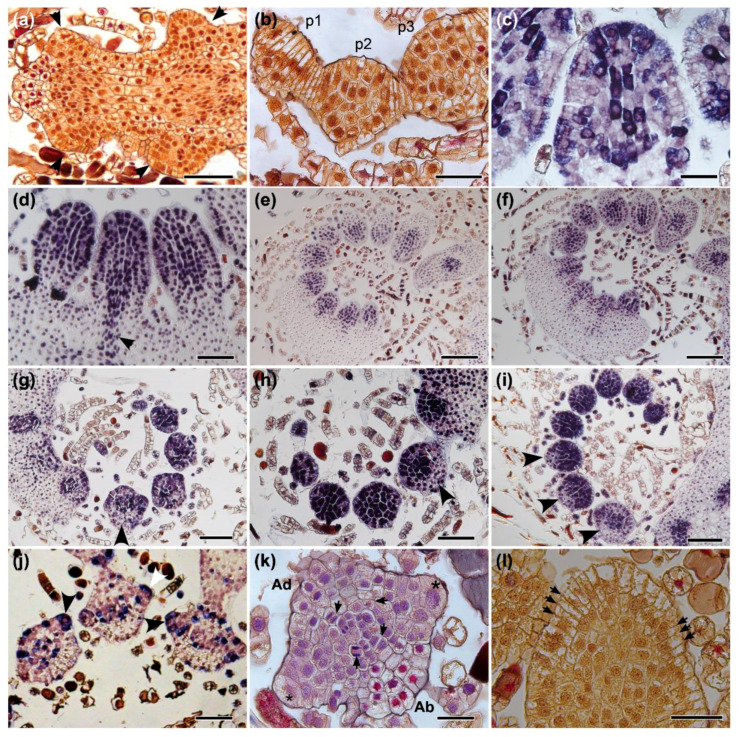
Late leaf development in terrestrial (Tf) and climbing (Cf) forms of Mickelia scandens by anatomical and expression analyses. Anatomical sections (a,b,k,l). (a) Pinnae primordia (arrowheads) emerge from the margins of the Tf leaf. (b) Transverse section of the youngest pinna (p1) shows grouped cells on its apex with evident periclinal divisions in Tf. The base has divisions in multiple planes, visible in older primordia (p2 and p3). In situ hybridization (c–j). (c) MsH4 expression indicates cells division in multiple adjacent cells at the apex of the pinna primordium and in the central axis, where the vasculature will develop in the Tf. (d) As the pinnae primordium increases in size, developing vascular traces express Class I KNOX genes, exemplified by MsC1KNOX1 in Cf. (e) MsC1KNOX1 in Cf and (f) MsC1KNOX2 in Cf are expressed in the entire young pinnae primordia. (g) MsH4 in Tf, (h) MsC1KNOX1 in Tf and (i) MsC1KNOX2 in Cf are all expressed throughout the entire pinnae primordia, expression is gradually reduced in the abaxial side of older pinnae (arrowheads). (j) Cell divisions detected by MsH4 expression in marginal cells, some indicated by arrows in Tf. (k) Anatomical transverse section of the pinna primordium showing marginal cells (*) with outer lenticular faces of the wall and submarginal initials, between adaxial (Ad) and abaxial (Ab) sides, and radial divisions in the center (arrows) in Cf. (l) Anatomical paradermal section of the pinna primordium showing rows of marginal cells, with anticlinal cutting faces, some indicated with arrows in Cf. Bars: (a,d,h,j) 100, (b,c,k,l) 50, (e,f) 200, (g,i) 150 µm.

