Figure 6.
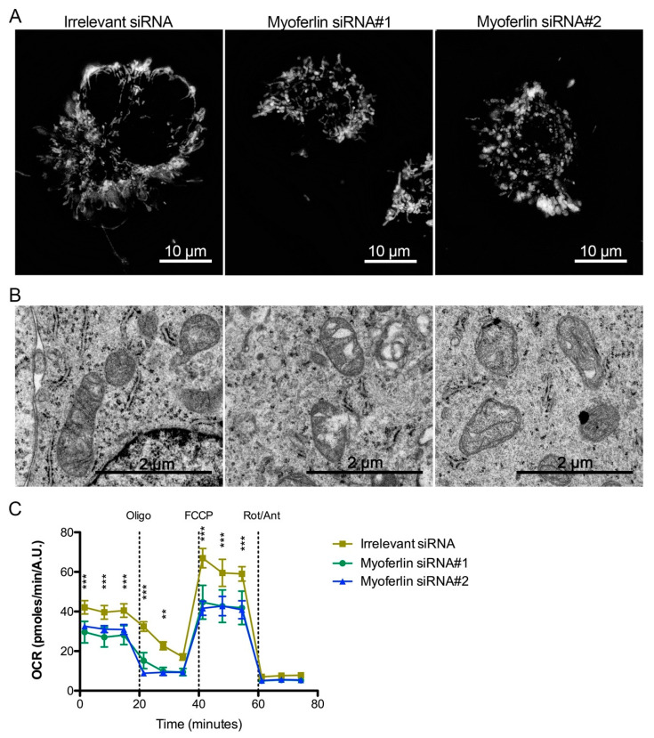
Mitochondrial impact of myoferlin deletion in pancreas cancer cells. (A) Mitochondria were stained with tetramethylrhodamine ethyl ester (1 nM TMRE) in Panc-1 living cells depleted for myoferlin. (B) Panc-1 cells depleted for myoferlin were fixed with glutaraldehyde and observed under transmission electron microscope. (C) Kinetic oxygen consumption rate (OCR) response of Panc-1 cells to oligomycin (oligo, 1 µM), FCCP (1.0 µM), rotenone, and antimycin A mix (Rot/Ant, 0.5 µM each). Each data point represents mean ± SD of technical replicates. All experiments were performed as three independent biological replicates. *** p < 0.001, ** p < 0.01.

