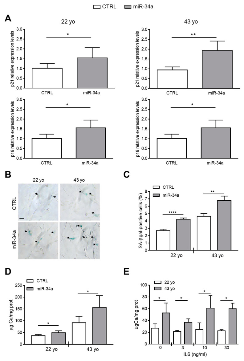
Figure 3.
Conditioned medium of miR-34a-overexpressing HASMCs enhances their senescence and calcification. Conditioned medium of HASMCs from 22- or 43-year-old (yo) donors was collected 72 h after infection with pMIRNA1 (CTRL) or pMIRH34a (miR-34a) lentivirus. (A) HASMCs were cultured for 24 h in the presence of corresponding conditioned medium. p21 and p16 expression was quantified by qRT-PCR and normalized to corresponding HPRT levels. Values are mean ± SD; *, p < 0.05; **, p < 0.01; Student’s t-test (22 yo); Mann–Whitney test (43 yo); n = 6. (B,C) HASMCs were cultured for 24 h in the presence of corresponding conditioned medium and processed for senescence-associated β-galactosidase (SA-β-gal) staining. (B) Representative images of SA-β-gal staining. Bar = 100 µm. (C) Bars show quantification of SA-β-gal-positive cells relative to (B). Values are mean ± SD; **, p < 0.01; ****, p < 0.0001; Student’s t-test; 22 yo, n = 3; 43 yo, n = 4. (D) Cells were cultured for 24 h in the presence of the corresponding conditioned medium and then in the osteogenic medium for 7 days. Calcification was measured by colorimetric analysis. Values are mean ± SD; *, p < 0.05; **, p < 0.01; ****, p < 0.0001; Student’s t-test; 22 yo, n = 6, 6; 43 yo, n = 6, 4. (E) HASMCs of 22- and 43 year-old (yo) donors were pretreated with indicated concentration of recombinant IL6 and subsequently cultured in osteogenic medium for 7 days. Calcification was measured by colorimetric analysis. Values are mean ± SD; *, p < 0.05; Student’s t-test; n = 3–4.

