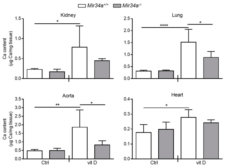
Figure 4.
Calcium deposition in Mir34a+/+ and Mir34a−/− mice after vitamin D treatment. Twelve-week-old Mir34a+/+ and Mir34a−/− mice were treated subcutaneously with either vitamin D (vit D) or a mock solution (Ctrl) for three consecutive days and sacrificed 5 days (Day 5) after the first injection. Calcium content in kidney, lung, heart and aorta (aortic arch) was quantified by the colorimetric analysis. Values are mean ± SD; *, p < 0.05; **, p < 0.01; ****, p < 0.0001; 1-way ANOVA followed by Bonferroni’s multiple comparison test; n = 5, 5, 4 and 5.

