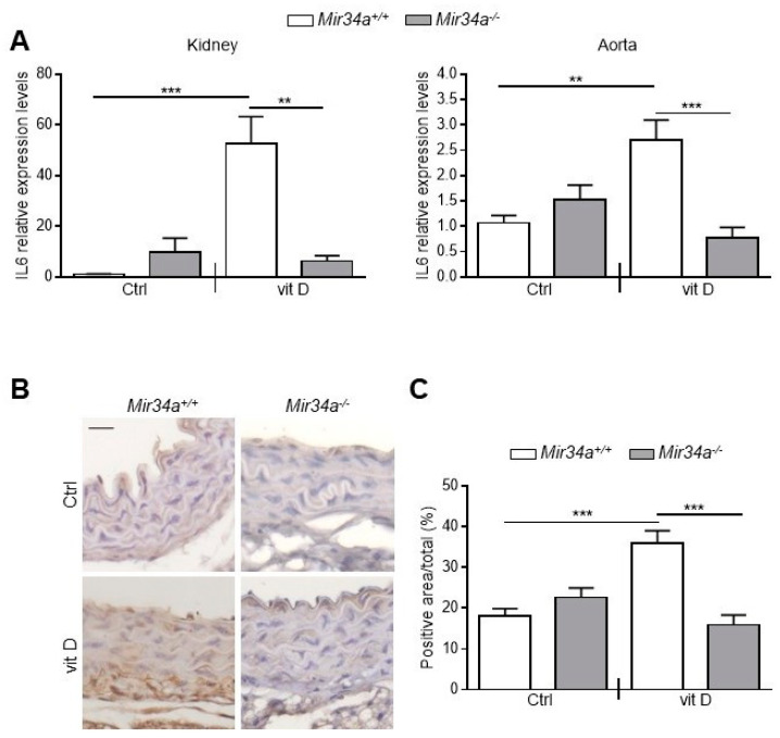
Figure 5.
IL6 expression in Mir34a+/+ and Mir34a−/− mice early after vitamin D treatment. Twelve-week-old Mir34a+/+ and Mir34a−/− mice were treated subcutaneously with either vitamin D (vit D) or a mock solution (Ctrl) for three consecutive days and sacrificed 3 days after the first injection (Day 3). (A) IL6 expression was analyzed by qRT-PCR and normalized to HPRT levels in the kidney and abdominal aorta. Values are mean ± SD. ** p < 0.01, *** p < 0.001; 1-way ANOVA with a Bonferroni post hoc test; n = 6, 5, 6–7 and 4–5. (B) Representative images of thoracic aorta sections stained for IL6 expression with a specific antibody. Bar = 20 μm. (C) Bars show quantification of the percentage of IL6 positive area to the total thoracic aortic area relative to (B). Values are mean ± SD; ***, p < 0.001; 1-way ANOVA followed by Bonferroni’s multiple comparison test; n = 4, 4, 4 and 3.

