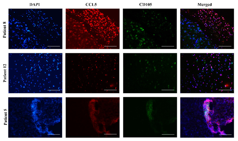
Figure 4.
Mesenchymal stem cells in glioblastoma tissues express CCL5. Fluorescence immunohistochemical staining of CCL5 antigen was performed on glioblastoma sections of 3 patients, Nb. 8, Nb. 12 and Nb. 5. MSCs were immunolabeled using the antibody against their specific marker CD105. Nuclei were stained with DAPI (blue), CCL5 with Alexa Fluor 546 (red), and CD105 with Alexa Fluor 488 (green) dye. Merged images represent colocalization (violet color) of CD105 and CCL5. Microscopy was carried out at 20× objective magnification. Scale bar represents 100 µm.

