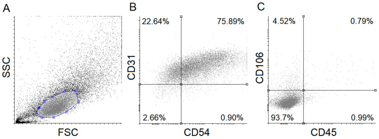
Figure 1.
Flow cytometric analysis of human umbilical cord vein endothelial cells (HUVECs). Isolated HUVECs were verified using specific fluorescent-labeled antibodies. Forward- and side-scatter plot and dot-plots (A) of HUVEC positive (CD54, CD31) (B) and negative (CD45, CD106) (C) markers are shown. FSC: Forward scatter, SSC: Side scatter.

