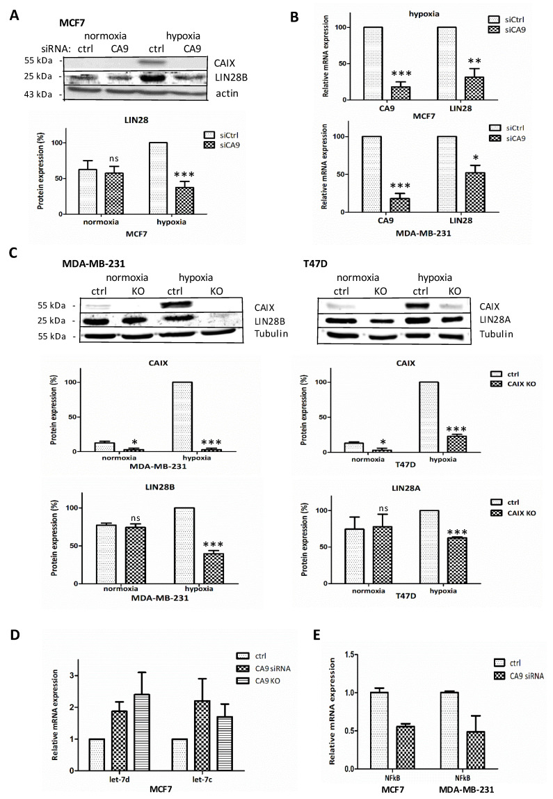
Figure 1.
CAIX suppression affects LIN28 expression. (A) Representative Western blot of normoxic and hypoxic MCF7 cells transiently transfected with siRNA-control/siRNA-CA9, detecting CAIX, actin, and LIN28. Signal intensities from three different immunoblots were quantified using the ImageJ software, normalized to actin, and presented as an average percentage of hypoxic control-siRNA (=100%). (B) Quantitative PCR analysis of CA9 and LIN28 mRNA expression in hypoxic CA9-silenced cells normalized to actin, presented as a percentage of the expression in cells transfected with control-siRNA (siCtrl = 100%). The results represent the mean of three independent biological experiments done in triplicates. (C) Representative Western blot of CAIX and LIN28 (homolog B for MDA-MB-231, homolog A for T47D) in MDA-MB-321-CA9-KO and T47D-CA9-KO cell lines. Average signal from 3 different Western blots quantified by the ImageJ software, normalized to tubulin, and presented as a percentage of hypoxic control (=100%). (D) The effect of CAIX-depletion on let-7 expression (qPCR) in MCF7, confirming microarray data presented in Table 1, extended with confirmation in KO approach. (E) NF-κB expression (qPCR) in si-CA9 suppressed MCF7 (according to microarray data presented in Table 1) and also in MDA-MB-231 cells. p > 0.05 was considered nonsignificant (ns), p < 0.05 is denoted as *, p < 0.01 as ** and p < 0.001 as ***.

