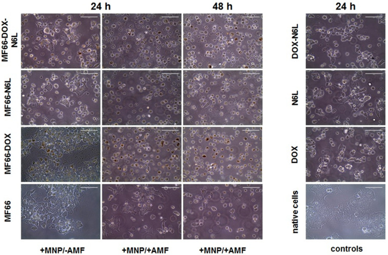
Figure 6.
Magnetic hyperthermia in combination with ((DOX and/or N6L) MNP formulations induced morphological changes and detachment of BT474 cells. Representative microscopic images of BT474 cells, which were incubated with the different MF66 formulations (100 µg Fe/mL, 24 h); 24 h or 48 h post magnetic hyperthermia (+MNPs/+AMF), MNPs control (+MNPs/−AMF), free agent controls (DOX, N6L or both) as well as untreated controls (native BT474). Scale bar = 100 µm.

