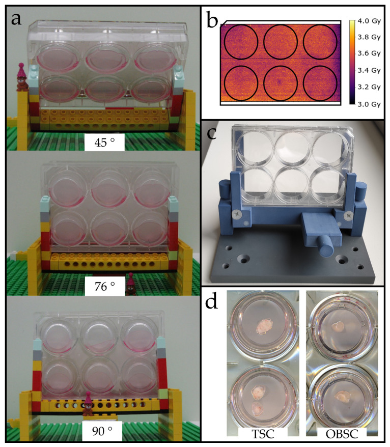
Figure 1.
Development and characterization of a proton irradiation setup for thin-cut tissue slices. (a) Exemplary tested angles using a Lego® rapid prototype. Membrane inserts tilted when angles >76° were used. Lower angels showed dose inhomogeneity. (b) Dose homogeneity for a 76° angle measured with EBT3 films on the plate bottom. (c) Adjustable milled setup at 76°. (d) Exemplary tumor (left) and brain slices (right) on the first day in culture.

