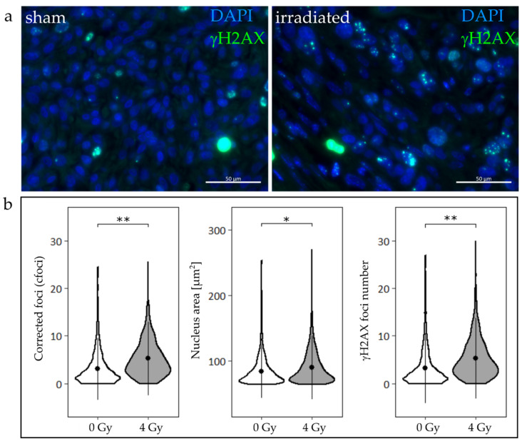
Figure 5.
Formation of γH2AX foci in tumor slices exposed to sham (0 Gy) or 4 Gy proton irradiation at 24 h post irradiation. (a) Representative immunofluorescent images of tumor slices treated with sham (left) or proton irradiation (right) show γH2AX foci. Apoptotic cells and endogenous DNA damages were observed in both groups; nevertheless, an increased number of foci could be detected in irradiated slices. (b) cfoci, nucleus area, and γH2AX foci numbers are significantly increased in slices that were irradiated with 4 Gy, compared to non-irradiated controls (linear mixed-effects model, *: p < 0.05, **: p < 0.01; nControl = 7, nTumor = 8).

