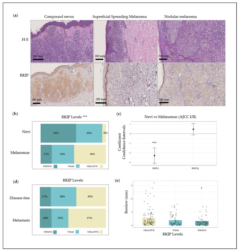
Figure 1.
Raf Kinase Inhibitor Protein (RKIP) expression in FFPE biopsies from patients. (a) Lower section of picture: IHC analysis of RKIP in normal melanocytes from a compound nevus, superficial spreading melanoma and nodular melanoma. Upper section of picture: Hematoxylin-Eosin staining (H-E); (b) RKIP staining distribution on nevus and melanoma tissue; (c) Coefficient confidence intervals for RKIP protein expression between nevus and melanoma samples; (d) RKIP staining distribution on histological sections of melanoma in stages I and II from patients who remained disease-free during follow-up versus patients who developed metastasis; (e) Kruskal–Wallis one-way analysis of Breslow index with respect to RKIP expression. * q < 0.05, *** q < 0.001.

