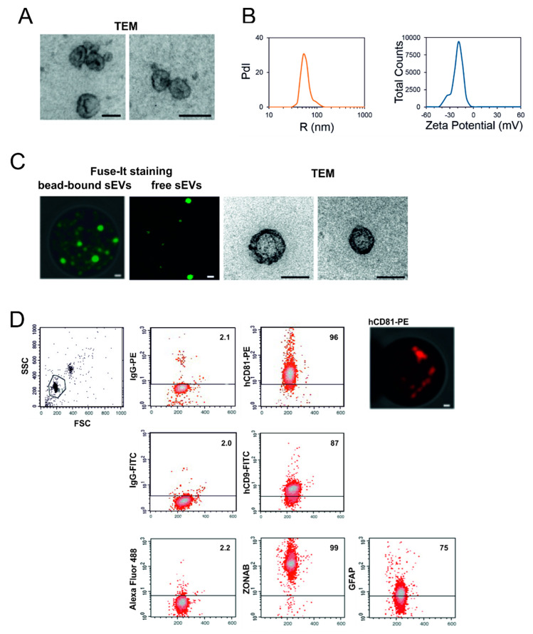
Figure 2.
Characterization of M14-released tumor-secreted small extracellular vesicles (sEVs) in cell culture. (A) Morphological examination of small extracellular vesicles (sEVs) purified from M14 cell culture medium was performed by transmission electron microscopy (TEM). Bars, 100 nm. (B) Size and number of the released sEVs was measured by dynamic light scattering. The representative Intensity distribution curve and Zeta potential distribution are an average of five different measurements of the same sample. (C) sEVs purified from cell culture were immunocaptured by magnetic Dynabeads conjugated with CD63 tetraspanin. The bead-bound sEVs stained by Fuse-It membrane-specific dye were studied by confocal microscopy (left panel, bars, 500 nm). The stained sEVs were then detached from the beads and analysed by confocal microscopy (middle panel, bars, 500 nm) and by TEM (right panels, bars, 100 nm). (D) Bead-bound sEVs were processed for the detection of the indicated molecules by immunofluorescence and flow cytometry. Aggregates and debris were excluded (gating) from the fluorescence analysis, as shown in the cytogram relative to the light scatter parameters (left panel, top). In each cytogram the number reported represents the percentage of positivity for the indicated molecule. As an example, right top panel reported the confocal microscopy of bead-bound sEVs stained with anti-CD81 antibody conjugated with phycoerythrin (PE). Bar, 500 nm. PdI, intensity distribution; SSC, side scatter; FSC, forward side scatter; FITC, fluorescein isothiocyanate; ZONAB, ZO-1-associated nucleic acid-binding protein; GFAP, glial fibrillary acidic protein.

